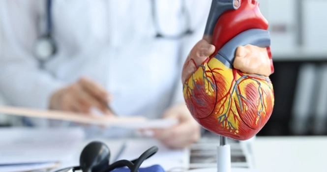
The Human Heart is central to our existence. It’s the first to start beating in the womb and the last one to stop at death.
Doctors who specialize in ailments or conditions related to the heart are,
- Cardiologists
- Cardiac Surgeon
Cardiologists are medical doctors who specialize in diagnosing, treating, and preventing diseases of the heart and blood vessels.
They undergo medical school and then complete a residency in internal medicine followed by a further specializing in cardiology.
Their focus is typically on non-surgical approaches to heart problems, although some subspecialists, such as interventional cardiologists, might perform minimally invasive procedures using catheters.
Cardiologists can further specialize in areas like pediatric cardiology, electrophysiology, heart failure, and more.
They work closely with patients to manage chronic conditions, provide preventive care, and coordinate with other healthcare professionals if surgical intervention is needed.
Cardiac surgeons, on the other hand, are specialists in performing surgical interventions on the heart and surrounding blood vessels.
They undergo medical school and then a residency in general surgery followed by a further specializing in cardiothoracic surgery.
Their training prepares them to perform complex operations such as heart transplants, coronary artery bypass grafting (CABG), heart valve repairs and replacements, and surgeries for congenital heart defects.
Cardiac surgeons work in close collaboration with cardiologists, especially when planning and performing surgeries, to ensure that the treatment is tailored to the specific needs and conditions of the patient.
After the surgery, the cardiac surgeon continues to monitor the patient’s recovery and may work alongside other healthcare professionals, including cardiologists, to manage postoperative care.
Let’s get to the basics and look into anatomy of the heart and conditions which require surgery.
Overview of Heart Structure
1. Chambers
The human heart consists of four chambers that pump blood throughout the body:
- Right Atrium: Collects oxygen-poor blood from the body and sends it to the right ventricle.
- Right Ventricle: Takes in deoxygenated blood from the right atrium and sends it to the lungs through the pulmonary artery.
- Left Atrium: Receives oxygen-rich blood from the lungs and pumps it into the left ventricle.
- Left Ventricle: Receives oxygenated blood from the left atrium and pumps it to the whole body through the aorta.
These chambers work in perfect coordination to sustain life by continuously circulating blood.
2. Valves
The heart contains four main valves that regulate blood flow between its chambers and to the rest of the body:
- Mitral Valve: Controls blood flow between the left atrium and left ventricle.
- Tricuspid Valve: Controls blood flow between the right atrium and right ventricle.
- Aortic Valve: Controls blood flow from the left ventricle into the aorta.
- Pulmonary Valve: Controls blood flow from the right ventricle into the pulmonary arteries.
These valves ensure that blood flows in the right direction, preventing backflow.
3. Vessels
The heart is connected to a complex network of blood vessels that carry blood to and from the body’s tissues:
- Aorta: The main artery that carries oxygenated blood from the left ventricle to the body.
- Pulmonary Arteries and Veins: Transport blood between the heart and lungs.
- Coronary Arteries: Supply oxygen-rich blood to the heart muscle itself.
- Vena Cava (Superior and Inferior): The main veins that return deoxygenated blood from the body to the right atrium.
These vessels create a closed circulatory system, facilitating the essential exchange of oxygen and nutrients with the body’s organs and tissues.
The intricate coordination of chambers, valves, and vessels in the heart enables it to fulfill its crucial role in sustaining life.
Any dysfunction within these components can lead to a cascade of health problems, making the understanding of their structure and function vital for both prevention and treatment of heart-related conditions.
Understanding Heart Function
1. Blood Circulation
The heart’s primary function is to pump blood throughout the body, ensuring the distribution of oxygen and nutrients to various organs and tissues, and the removal of waste products like carbon dioxide. Blood circulation occurs in two main pathways:
- Systemic Circulation: Oxygenated blood is pumped from the left ventricle into the aorta, traveling through arteries, arterioles, and capillaries to reach the body’s tissues. Deoxygenated blood returns via veins to the right atrium.
- Pulmonary Circulation: Deoxygenated blood is pumped from the right ventricle to the lungs through the pulmonary arteries. In the lungs, it picks up oxygen and releases carbon dioxide before returning to the left atrium through the pulmonary veins.
2. Heartbeat Regulation
The heartbeat is orchestrated by the heart’s electrical system:
- Sinoatrial (SA) Node: Often referred to as the heart’s natural pacemaker, the SA node initiates each heartbeat, sending an electrical impulse that causes the atria to contract.
- Atrioventricular (AV) Node: The electrical signal travels to the AV node, then down the Bundle of His and into the Purkinje fibers, causing the ventricles to contract.
- Autonomic Nervous System: The sympathetic and parasympathetic nervous systems can modulate the heart rate and force of contraction in response to the body’s needs, such as during exercise or stress.
3. Valve Function
As mentioned previously, the heart’s valves ensure unidirectional blood flow:
- Atrioventricular Valves (Mitral and Tricuspid): Prevent backflow into the atria when the ventricles contract.
- Semilunar Valves (Aortic and Pulmonary): Prevent backflow into the ventricles when the heart relaxes.
4. Nutrient and Waste Exchange
The heart’s pumping action facilitates the exchange of nutrients and waste products at the capillary level throughout the body. This process sustains cell function and overall body homeostasis.
5. Coronary Circulation
The heart itself requires a constant supply of oxygen and nutrients to function effectively. The coronary arteries provide this supply, and any blockage can lead to severe problems, such as a heart attack.
6. Hormonal and Pressure Regulation
The heart also plays a role in regulating blood pressure and producing hormones like atrial natriuretic peptide (ANP), which affects salt and water balance.
Conditions that Require Heart Surgery
Coronary Artery Disease (CAD)
Coronary Artery Disease is one of the most common heart conditions that may necessitate surgical intervention. It’s characterized by the narrowing or blockage of the coronary arteries, which are responsible for supplying the heart muscle with oxygen-rich blood.
Causes:
CAD typically results from a buildup of fatty deposits (atherosclerosis) in the arteries. Other contributing factors include high blood pressure, high cholesterol, smoking, diabetes, obesity, and a sedentary lifestyle.
Symptoms:
The disease might be asymptomatic in its early stages. As it progresses, symptoms can include chest pain (angina), shortness of breath, fatigue, weakness, and even heart attack if a complete blockage occurs.
Treatment and Surgical Interventions:
- Medications and Lifestyle Changes: These are often the first line of treatment, focusing on reducing risk factors and managing symptoms.
- Angioplasty and Stent Placement: Performed by interventional cardiologists, this procedure opens narrowed arteries and can include the placement of a stent to keep the artery open.
- Coronary Artery Bypass Grafting (CABG): Conducted by cardiac surgeons, CABG creates new pathways around blocked arteries using veins or arteries from other parts of the body. It’s often recommended for severe cases of CAD or multiple blockages.
Prognosis:
With proper treatment and lifestyle modifications, many individuals with CAD can lead a normal life. However, it is a chronic condition that requires ongoing medical care and monitoring.
Coronary Artery Disease is a prevalent condition that significantly impacts heart health. Depending on its severity and the patient’s overall health, various interventions, including surgical options, might be necessary.
Prompt diagnosis and appropriate treatment are vital in managing CAD effectively, as they can prevent further complications and improve the quality of life for those affected.
Valve Disease
Valve Disease refers to any dysfunction or disorder of one or more of the heart’s four valves: the mitral, tricuspid, aortic, and pulmonary valves. These valves control the direction of blood flow through the heart’s chambers, and any malfunction can have serious consequences.
Types:
Valve diseases can be categorized mainly into:
- Stenosis: Narrowing of a valve, restricting blood flow.
- Regurgitation: Incomplete closure of a valve, leading to backward flow.
- Atresia: A valve lacks an opening for blood to pass through.
- Prolapse: A valve doesn’t close properly, often because of weakened supportive tissues.
Causes:
Causes can be congenital (present at birth) or acquired later in life, often due to:
- Aging
- Infections like rheumatic fever
- Heart attack or injury to the heart
- Connective tissue disorders
- Radiation therapy to the chest
Symptoms:
Symptoms may vary widely and can be nonexistent or vague, especially in the early stages. They might include:
- Fatigue
- Shortness of breath
- Chest pain or palpitations
- Swelling in the ankles, feet, or abdomen
- Lightheadedness or fainting
Treatment and Surgical Interventions:
Treatment depends on the type, cause, and severity of the valve disease:
- Medications: To manage symptoms and prevent further damage.
- Balloon Valvuloplasty: A catheter with a balloon is used to widen a stenotic valve.
- Valve Repair: Preserving the patient’s own valve and repairing it.
- Valve Replacement: Replacing the diseased valve with a prosthetic one.
Surgical options, particularly repair or replacement, are often recommended for moderate to severe cases.
Prognosis:
With appropriate treatment, many people with valve disease can lead normal, active lives. Regular follow-up care is essential to monitor the condition and make necessary treatment adjustments.
Aortic Aneurysm
An aortic aneurysm is an abnormal bulge that occurs in the wall of the aorta, the largest blood vessel in the body that carries oxygenated blood from the heart to the rest of the body. Depending on its size and location, an aortic aneurysm can be a serious health risk.
Types:
Aortic aneurysms can be classified into:
- Thoracic Aortic Aneurysm: Occurs in the chest region.
- Abdominal Aortic Aneurysm: Occurs in the abdominal area.
Causes:
The exact cause can be unknown, but common factors include:
- Atherosclerosis (hardening of the arteries)
- High blood pressure
- Genetic factors or familial history
- Infections or trauma
Symptoms:
Many aortic aneurysms are asymptomatic and may be discovered during an examination for another condition. If symptoms do appear, they may include:
- Pain in the chest, back, or abdomen
- Shortness of breath
- Coughing or hoarseness
- Difficulty swallowing
Risks:
The primary risk is rupture, leading to life-threatening internal bleeding. The risk increases with the size of the aneurysm
Treatment and Surgical Interventions:
Treatment depends on the size, type, and location of the aneurysm:
- Monitoring: Small aneurysms might only require regular monitoring.
- Medications: To manage blood pressure and cholesterol.
- Endovascular Repair: A minimally invasive procedure using a stent-graft.
- Open Surgical Repair: Replacement or reinforcement of the affected aorta segment with a synthetic graft.
Surgery is typically recommended for larger aneurysms or those that grow quickly, as these pose a higher risk of rupture.
Prognosis:
With early diagnosis and appropriate treatment, the prognosis for an aortic aneurysm can be favorable. However, a ruptured aneurysm has a high mortality rate and requires emergency intervention.
Heart Failure
Heart failure is a chronic, progressive condition in which the heart muscle is unable to pump blood efficiently enough to meet the body’s needs for blood and oxygen. It doesn’t mean the heart has stopped working, but rather that it’s not working as well as it should.
Types:
Heart failure can be categorized into:
- Left-Sided Heart Failure: The left ventricle doesn’t pump blood efficiently, causing fluid to back up into the lungs.
- Right-Sided Heart Failure: Often a result of left-sided failure, leading to fluid accumulation in the abdomen, legs, and other peripheral tissues.
- Congestive Heart Failure: A combination of both, leading to generalized fluid retention.
Causes:
The condition may result from various underlying factors, including:
- Coronary artery disease
- High blood pressure
- Faulty heart valves
- Myocarditis (inflammation of the heart muscle)
- Cardiomyopathies (diseases of the heart muscle)
- Diabetes
Symptoms:
Symptoms may vary based on the severity and type of heart failure, commonly including:
- Shortness of breath
- Persistent coughing or wheezing
- Fatigue or weakness
- Swelling in legs, ankles, and feet
- Rapid weight gain due to fluid retention
- Lack of appetite or nausea
Treatment:
Heart failure is a lifelong condition but can be managed through:
- Lifestyle Changes: Including diet, exercise, weight management, and avoiding alcohol and smoking.
- Medications: To improve heart function, reduce symptoms, and prevent further damage.
- Devices and Surgical Procedures: Such as implantable cardioverter-defibrillators (ICDs), cardiac resynchronization therapy (CRT), or heart pumps.
In severe cases, a heart transplant may be considered.
Prognosis:
While heart failure is a serious condition, many people are able to manage it with proper treatment and lifestyle adjustments. Regular medical follow-up is crucial for ongoing care and adjustment of treatments.
Arrhythmias
Arrhythmias refer to any disorder of the heart rate or rhythm. They occur when the electrical impulses that coordinate heartbeats are not functioning properly, leading to heartbeats that are too fast, too slow, or irregular.
Types:
Arrhythmias are classified into several categories, including:
- Bradycardia: A heart rate that’s too slow (Less than 60 beats per minute)
- Tachycardia: A heart rate that’s too fast (More than 100 beats per minute)
- Atrial Fibrillation (AFib): A common type, characterized by rapid and irregular beating in the atria.
- Ventricular Fibrillation (VFib): A serious condition where the ventricles quiver instead of pumping effectively.
Causes:
Several factors can contribute to arrhythmias, including:
- Heart disease
- High blood pressure
- Electrolyte imbalances
- Infections or fever
- Genetic predisposition
- Substance abuse (including alcohol and certain medications)
Symptoms:
Some arrhythmias might be asymptomatic, while others may cause symptoms such as:
- Palpitations or feeling of skipped beats
- Dizziness or lightheadedness
- Fainting (syncope)
- Shortness of breath
- Chest pain
Diagnosis:
Various diagnostic tests can detect arrhythmias, including:
- Electrocardiogram (ECG): To record electrical signals.
- Holter Monitor: A portable ECG device worn for a day or more.
- Event Monitor: A device to record specific episodes.
- Stress Testing: To observe heart rhythm under exertion.
Treatment:
Treatment for arrhythmias may include:
- Lifestyle Changes: Such as healthy diet, exercise, and avoiding triggers like caffeine or alcohol.
- Medications: To control heart rate or rhythm.
- Catheter Ablation: To destroy abnormal tissue causing the arrhythmia.
- Pacemaker or ICD Implantation: To regulate heart rhythm.
- Surgery: In some severe cases, surgery may be necessary.
Prognosis:
The outlook for arrhythmias varies widely depending on the type, cause, and overall heart health. Many are manageable with treatment, while others can be life-threatening without intervention.
Congenital Heart Defects
Congenital heart defects (CHDs) are abnormalities in the heart’s structure that are present at birth. They represent a diverse group of disorders that can range from simple to complex and may affect the heart’s walls, valves, arteries, and veins.
Types:
There are many types of congenital heart defects, including:
- Septal Defects: Holes in the heart’s septum, like atrial septal defect (ASD) or ventricular septal defect (VSD).
- Valve Defects: Abnormalities in one or more heart valves, such as stenosis or atresia.
- Tetralogy of Fallot: A combination of four defects leading to deoxygenated blood flowing into the body.
- Coarctation of the Aorta: Narrowing of the aorta that can restrict blood flow.
- Transposition of the Great Arteries: A condition where the main arteries are switched.
Causes:
The exact cause of CHDs is often unknown, but they may be related to:
- Genetic factors and familial history.
- Environmental factors, such as maternal exposure to certain medications, alcohol, or infections during pregnancy.
- Underlying maternal health conditions, like diabetes.
Symptoms:
Symptoms vary widely based on the defect and can range from mild to severe. They might include:
- Blue-tinged skin, lips, or fingernails (cyanosis)
- Difficulty breathing or feeding
- Fatigue
- Swelling in the legs, abdomen, or areas around the eyes
Diagnosis:
Diagnosis can occur before birth, shortly afterward, or even later in life. Tools for diagnosis include:
- Prenatal Ultrasound: Can sometimes detect defects in utero.
- Physical Examination: To identify symptoms such as a heart murmur.
- Echocardiogram: To provide detailed images of the heart.
- Cardiac Catheterization: To assess the heart’s internal structure and function.
Treatment:
Treatment depends on the type and severity of the defect and may include:
- Monitoring and Medications: For milder defects.
- Catheter Procedures: To repair holes or open narrowed vessels.
- Surgery: To repair complex defects, sometimes requiring multiple operations over years.
Prognosis:
The prognosis for CHDs has improved dramatically with advancements in diagnostics and treatment. Many individuals with CHDs lead healthy lives, although they may require ongoing care and monitoring.
Procedures Performed by Cardiologists
1. Angiography:
Utilizes X-rays to visualize blood flow in the coronary arteries.
Angiography is a medical imaging technique used to visualize the blood vessels within various body regions, including the heart. It is particularly valuable in diagnosing and managing heart conditions, such as coronary artery disease (CAD).
During the procedure, a contrast dye is injected into the blood vessels, and X-ray images are taken to provide a detailed view of the arteries and veins.
In cardiac angiography, this allows physicians to detect blockages, narrowings, or other abnormalities in the coronary arteries that might require intervention, such as angioplasty or bypass surgery.
Angiography can also help assess the blood flow to the heart muscles and the function of the heart valves.
While it is an essential tool in cardiovascular diagnosis and treatment, angiography is an invasive procedure and carries some risks, such as bleeding, infection, or allergic reaction to the contrast dye.
Proper care and medical supervision are necessary to minimize these risks, ensuring that angiography remains a vital and effective tool in heart care.
2. Angioplasty and Stent Placement:
Expands narrowed or blocked arteries and may include the placement of a stent to keep the artery open.
Angioplasty and stent placement are common procedures used to open narrowed or blocked blood vessels, typically in the context of coronary artery disease (CAD).
Angioplasty:
- Procedure: Angioplasty involves threading a catheter with a small inflatable balloon at its tip through the blood vessels to the affected artery. Once in place, the balloon is inflated, compressing the plaque against the artery walls and widening the vessel to restore blood flow.
- Use: It’s primarily used to relieve symptoms of heart disease or reduce damage during or after a heart attack.
Stent Placement:
- Procedure: Often performed in conjunction with angioplasty, stent placement involves inserting a small wire mesh tube called a stent into the newly opened artery. The stent helps keep the artery open, reducing the chance of it narrowing again.
- Types of Stents: Drug-eluting stents release medication to prevent the artery from closing again. Bare-metal stents do not have this coating.
- Use: Stents are used to reduce symptoms, lower the risk of heart attacks, and help improve the prognosis in patients with heart disease.
Benefits:
- Minimally Invasive: Both angioplasty and stent placement are less invasive than open-heart surgery.
- Short Recovery Time: Most patients can return home within a day or two and resume normal activities shortly afterward.
- Effective Symptom Relief: These procedures can significantly relieve symptoms like chest pain and shortness of breath.
Risks and Considerations:
- Complications: Risks include bleeding, infection, clotting within the stent, or re-narrowing of the artery (restenosis).
- Ongoing Care: Patients receiving stents usually require long-term medication to prevent blood clots and regular follow-up appointments.
3. Electrophysiology Study (EPS):
Examines electrical activity in the heart to diagnose arrhythmias.
An Electrophysiology Study (EPS) is a specialized procedure that examines the electrical conduction system of the heart. This diagnostic test is vital in understanding the nature of abnormal heart rhythms or arrhythmias.
During an EPS, catheters equipped with electrodes are threaded into the heart from a blood vessel in the groin, arm, or neck.
These catheters can both record the heart’s electrical activity and artificially stimulate the heart to induce various rhythms.
This dual capability allows healthcare providers to pinpoint the origin of an arrhythmia and analyze how electrical signals travel through the heart.
The EPS can help in determining the most effective treatment approach, be it medication, a pacemaker, catheter ablation, or other interventions.
While an invaluable tool in the management of heart rhythm disorders, the procedure is complex and must be conducted with great care to avoid potential risks such as bleeding, infection, or damage to the heart or surrounding structures.
4. Catheter Ablation:
Treats arrhythmias by destroying the heart tissue responsible for the irregular rhythm.
Catheter Ablation is a medical procedure used to treat certain types of arrhythmias, or abnormal heart rhythms.
It involves guiding a catheter (a thin, flexible tube) to the heart through the blood vessels, typically from the groin or neck.
Once the specific area of abnormal electrical activity is identified, usually through a preceding Electrophysiology Study (EPS), the tip of the catheter emits energy (such as radiofrequency energy) to create a small scar in the heart tissue.
This scar interrupts the abnormal electrical pathway, restoring normal heart rhythm. Catheter ablation has become a preferred treatment for many arrhythmias, including atrial fibrillation and supraventricular tachycardia, as it offers a potentially curative option with less invasiveness compared to open-heart surgery.
Success rates are generally high, especially for certain types of arrhythmias, but risks include possible damage to the heart or blood vessels, bleeding, or infection. Careful patient selection, expert procedural execution, and appropriate follow-up are essential to optimize outcomes.
5. Echocardiography:
Utilizes ultrasound to create images of the heart’s structure and function.
Echocardiography, commonly referred to as an echocardiogram or “echo,” is a non-invasive ultrasound imaging technique used to visualize the heart’s structure and function.
By using sound waves, it produces detailed images of the heart’s chambers, valves, walls, and blood vessels. Echocardiography can reveal important information about the size and shape of the heart, the function of the heart’s pumping chambers (ventricles), the operation of its valves, and the flow of blood within it.
It is widely used to diagnose and manage various heart conditions, ranging from heart failure and congenital heart disease to valve disorders and cardiac masses.
Echocardiography also plays a crucial role in monitoring heart health following surgeries or other treatments. The procedure is typically performed by a sonographer or cardiologist and is considered very safe, without the risks associated with radiation or invasive techniques.
Its accessibility, non-invasive nature, and ability to provide real-time images make echocardiography an indispensable tool in modern cardiology.
6. Cardiac Stress Testing:
Measures the heart’s response to stress, often through exercise or medication.
Cardiac Stress Testing, often simply referred to as a stress test, is a diagnostic procedure used to assess how well the heart works under physical stress.
During the test, patients are asked to exercise, usually on a treadmill or stationary bicycle, while their heart rate, blood pressure, and electrocardiogram (ECG) are monitored.
In cases where physical exercise is not suitable, medications can be administered to simulate the effect of exercise on the heart.
The primary goal of the stress test is to identify any abnormal blood flow to the heart’s muscle tissue, which may indicate underlying coronary artery disease (CAD).
It can also help determine the patient’s exercise tolerance, the effectiveness of cardiac treatments, and the risk of future cardiac events.
Cardiac Stress Testing is widely utilized in cardiology to provide valuable insights but must be performed under careful medical supervision to manage potential risks and ensure accurate interpretation of the results. It forms an integral part of the diagnostic and risk-assessment toolkit for managing heart disease.
7. Pacemaker and Defibrillator Implantation:
Involves implanting devices to regulate heart rhythm.
Pacemaker and Defibrillator Implantation are life-saving procedures that involve the placement of small devices to regulate the heart’s rhythm.
A pacemaker is a device designed to control slow heart rhythms by sending electrical impulses to stimulate the heart when necessary. It’s typically used in conditions like bradycardia, where the heart beats too slowly.
A defibrillator, on the other hand, monitors the heart for dangerously fast rhythms, like ventricular fibrillation, and delivers a shock to reset the heart’s normal rhythm when needed.
Both devices are implanted under the skin, usually near the collarbone, and connected to the heart through one or more leads. Implantation is performed under local anesthesia, and the devices are programmed to meet individual patient needs.
These procedures are essential for many patients with heart rhythm disorders, allowing them to live normal lives with reduced risk of sudden cardiac events.
While implantation is generally safe and effective, potential risks include infection, bleeding, or issues with the leads, and regular follow-up is required to ensure proper functioning of the devices.
8. Transesophageal Echocardiogram (TEE):
Provides a close look at the heart’s structure and function through the esophagus.
Transesophageal Echocardiogram (TEE) is an advanced form of echocardiography that offers detailed images of the heart and its structures from a unique vantage point.
Unlike the standard echocardiogram, where the ultrasound probe is placed on the chest’s surface, TEE involves inserting a specialized probe down the patient’s esophagus.
Given the esophagus’s proximity to the heart, TEE provides exceptionally clear, high-resolution images, especially of structures like the heart’s back chambers, valves, and the aorta.
This procedure is invaluable in detecting conditions such as infections on the heart valves (endocarditis), congenital heart defects, and blood clots in the heart’s chambers.
As the probe is introduced through the mouth and into the esophagus, patients are usually sedated to ensure comfort during the procedure. While TEE is generally safe, care must be taken to avoid complications such as esophageal injury or reactions to sedation.
The unparalleled clarity and detail offered by TEE make it a critical tool in the diagnostic arsenal of cardiologists and cardiac surgeons.
Procedures performed by Cardiac Surgeons
1. Coronary Artery Bypass Grafting (CABG):
Redirects blood flow around blocked coronary arteries.
Coronary Artery Bypass Grafting (CABG) is a surgical procedure commonly referred to as heart bypass surgery, designed to improve blood flow to the heart muscle.
In individuals with significant blockages in their coronary arteries due to plaque buildup, the heart can be deprived of oxygen-rich blood, leading to chest pain (angina) or heart attacks.
During CABG, a surgeon takes a blood vessel from another part of the body—often the chest, leg, or arm—and uses it to create a new route around the blocked artery, effectively bypassing the obstruction.
This “graft” restores the flow of blood, supplying the heart muscle with the essential oxygen and nutrients it needs to function effectively. CABG can involve bypassing multiple arteries, depending on the extent of coronary artery disease.
The procedure has been practiced for decades and has been instrumental in saving countless lives. While CABG is invasive and requires a recovery period, it offers a long-term solution to severe coronary artery blockages and can significantly improve the patient’s quality of life and longevity.
2. Heart Valve Repair or Replacement:
Repairs or replaces malfunctioning heart valves.
Heart Valve Repair or Replacement surgery addresses malfunctions in one or more of the heart’s four valves: the aortic, mitral, pulmonary, or tricuspid.
These valves ensure unidirectional blood flow through the heart, and when they don’t function correctly, it can lead to conditions like stenosis (narrowing of the valve) or regurgitation (backward flow of blood due to a leaky valve).
The choice between repair or replacement typically depends on the valve’s condition and the extent of damage. Valve repair often involves modifying the original valve to restore its function, using procedures like ring annuloplasty (where a ring is sewn around the base of the valve to tighten it) or commissurotomy (separating fused valve leaflets).
When repair isn’t viable, a replacement is done using mechanical valves, made from materials like titanium or carbon, or biological valves, derived from pig, cow, or human donors.
The decision on the type of valve used factors in patient age, medical condition, and lifestyle. Both repair and replacement procedures are major surgeries that can significantly improve or prolong life, but they come with risks and may require lifelong medication, especially with mechanical valves, to prevent complications like blood clots.
3. Surgical Treatment for Arrhythmias:
Includes procedures like the Maze surgery to correct abnormal heart rhythms.
Surgical Treatment for Arrhythmias addresses the irregular heart rhythms that can compromise the heart’s efficiency and overall health.
Arrhythmias, originating from problems in the heart’s electrical system, can range from benign to life-threatening. While many arrhythmias are managed with medication, catheter ablation, or devices like pacemakers, some severe cases necessitate surgical intervention.
One such procedure is the Maze surgery, particularly effective for atrial fibrillation. During this operation, a series of surgical incisions are made in the atria (the heart’s upper chambers), which are then stitched together.
As they heal, scar tissue forms, creating a “maze” that prevents the abnormal electrical signals causing the arrhythmia from spreading. Another surgical option is the implantation of an Automatic Implantable Cardioverter-Defibrillator (AICD), a device that continuously monitors heart rhythm and delivers electric shocks when necessary to restore a regular heartbeat.
While surgical treatments for arrhythmias can be highly effective, they are typically reserved for cases where other less invasive treatments have failed or are deemed unsuitable.
4. Aortic Aneurysm Repair:
Repairs weakened and bulging areas in the aorta.
Aortic Aneurysm Repair is a critical surgical intervention aimed at treating an aortic aneurysm, which is a bulging or dilation of a section of the aorta, the body’s main artery.
If left untreated, the aneurysm can rupture, leading to life-threatening internal bleeding. The repair procedure chosen largely depends on the size, location, and shape of the aneurysm.
There are two primary approaches to this repair: open surgery and endovascular repair.
Open surgery involves making a large incision in the chest or abdomen to access the aorta, removing the dilated section, and replacing it with a synthetic graft.
Endovascular aneurysm repair (EVAR), on the other hand, is less invasive and involves threading a catheter through the femoral artery in the groin, guiding it to the aneurysm site, and placing a stent-graft to reinforce the weakened segment of the aorta.
While EVAR offers a quicker recovery and fewer immediate risks, it may require regular follow-up and has a chance of future interventions.
The decision between these methods is made after weighing the risks and benefits, considering the patient’s overall health, and the specific characteristics of the aneurysm. Regardless of the approach, timely repair is crucial to prevent potentially catastrophic outcomes.
5. Left Ventricular Assist Device (LVAD) Implantation:
Implants a mechanical device to support heart function and blood flow in patients with heart failure.
Left Ventricular Assist Device (LVAD) Implantation involves the placement of a mechanical pump that aids a weakened or failing left ventricle, the heart’s main pumping chamber. An LVAD doesn’t replace the heart, but instead supports its function by taking over or supplementing its pumping capability.
It is primarily used for patients with advanced heart failure, serving two main purposes: as a “bridge to transplant” for those awaiting a heart transplant or as a “destination therapy” for those who aren’t transplant candidates but need long-term heart function support.
The device comprises a pump placed inside the body, connected to the heart, with a driveline exiting the body and attaching to an external controller and power sources.
Over the years, advancements in LVAD technology have resulted in smaller, more efficient devices with fewer complications.
While LVAD implantation can significantly improve quality of life and survival rates, patients and caregivers need thorough education about living with the device, especially concerning maintenance, potential complications, and emergency procedures.
The procedure is a testament to the remarkable strides made in treating advanced heart disease and offers hope to many individuals with previously limited options.
6. Congenital Heart Defect Repairs:
Surgical correction of heart defects present at birth.
Congenital Heart Defect Repairs refer to a spectrum of surgical procedures aimed at correcting heart anomalies present at birth. These defects, ranging from simple to complex, can affect the heart’s structure, including its walls, valves, and major blood vessels.
Simple defects, like small holes in the heart’s chambers (atrial or ventricular septal defects), might be closed using cardiac catheterization techniques or open-heart surgery.
More complex defects, such as Tetralogy of Fallot or transposition of the great arteries, necessitate intricate surgical repairs or even multiple staged operations over time. The primary goal of these procedures is to improve circulation, optimize heart function, and prevent complications like heart failure or arrhythmias.
Advancements in diagnostic techniques, surgical methods, and post-operative care over the decades have drastically improved the outcomes of these repairs. Today, due to these advancements, many children with congenital heart defects grow into adulthood and lead active, fulfilling lives. However, lifelong medical follow-up is often essential, as some repairs might require adjustments or reinterventions in the future.
7. Transmyocardial Laser Revascularization (TMLR):
Creates channels in the heart muscle to improve blood flow to the heart’s chambers.
Transmyocardial Laser Revascularization (TMLR) is a surgical procedure developed as an alternative treatment for individuals with severe angina (chest pain) who aren’t candidates for traditional interventions like coronary artery bypass grafting (CABG) or angioplasty.
Patients with diffuse coronary artery disease often have areas of the heart muscle that don’t receive adequate blood flow, leading to debilitating angina. TMLR aims to improve this blood flow.
During the procedure, a surgeon uses a laser to create a series of small channels in the heart muscle, specifically in the left ventricle. While the exact mechanism of relief isn’t entirely clear, it’s believed that these channels promote the growth of new blood vessels (angiogenesis) and possibly also redirect blood flow within the heart muscle.
Additionally, TMLR may reduce pain by destroying nerve endings in the heart muscle. While it doesn’t replace conventional treatments, TMLR has shown promise in reducing angina and improving the quality of life for specific subsets of patients, especially when combined with other therapies. It remains a specialized procedure, offered at select cardiac centers to those who have exhausted other treatment options.
8. Heart Transplant:
Replaces a failing heart with a healthy donor heart.
A heart transplant is a life-saving surgical procedure in which a diseased or failing heart is replaced with a healthy donor heart. It is considered the final therapeutic option for patients with end-stage heart failure or certain severe heart conditions when all other treatments have failed.
The process begins with a thorough evaluation to determine if a patient is a suitable candidate, taking into account both medical and psychosocial factors.
Once approved, the patient is placed on a waiting list, with organ allocation typically based on urgency, compatibility, and waiting time. The procedure itself is complex, requiring the recipient’s heart to be removed (with the exception of some bi-atrial techniques where parts of the atria remain) and the donor heart to be connected to the major blood vessels.
Post-operative care is critical, with patients receiving immunosuppressive drugs to prevent organ rejection—a condition where the body’s immune system attacks the new heart.
These medications, while essential, can come with side effects and increase susceptibility to infections. Regular medical follow-ups are imperative to monitor heart function, detect potential complications, and ensure optimal health.
Over the years, advancements in surgical techniques, post-operative care, and immunosuppressive therapies have improved survival rates and quality of life for transplant recipients. Nevertheless, a heart transplant remains a profound experience, with recipients often requiring lifelong medical care and emotional support.
Diagnostic Tests Prior to Heart Surgery
Heart surgery is a significant medical procedure, and several diagnostic tests are usually performed beforehand to assess the heart’s function and structure, as well as to identify any potential risks. These tests help in planning the surgery and anticipating possible complications.
A. Electrocardiogram (EKG)
An Electrocardiogram (EKG or ECG) is a non-invasive diagnostic test that records the electrical activity of the heart over a period of time. It is commonly used prior to heart surgery for several reasons:
1. Assessing Heart Rhythm and Rate:
An EKG can detect abnormal heart rhythms (arrhythmias), such as atrial fibrillation or ventricular tachycardia, which may need to be addressed before surgery.
2. Detecting Heart Disease:
Changes in the EKG pattern can signal underlying heart disease, previous heart attacks, or other cardiac problems, providing valuable information about the heart’s condition.
3. Guiding Surgical Planning:
Understanding the heart’s electrical patterns can help surgeons plan the procedure more accurately, particularly if the surgery is related to arrhythmias or other electrical problems within the heart.
4. Baseline Measurement:
Performing an EKG before surgery establishes a baseline for the patient’s heart activity. This can be compared to future EKGs to detect any changes or problems that may develop postoperatively.
5. Identifying Potential Risks:
Certain EKG findings may indicate increased risk during surgery, such as ischemia or severe electrolyte imbalances. Identifying these risks ahead of time allows for necessary adjustments in surgical planning and anesthesia.
Procedure:
The EKG is performed by attaching electrodes to the skin on the chest, arms, and legs. These electrodes are connected to a machine that translates the electrical signals into wave patterns on a graph. The procedure is painless and typically takes only a few minutes.
B. Echocardiogram
An echocardiogram, often referred to simply as an “echo,” is a diagnostic test that utilizes ultrasound technology to create detailed images of the heart’s structure and function. It’s a vital tool used before heart surgery for several essential reasons:
1. Assessing Heart Structure:
The echocardiogram provides a visual representation of the heart’s chambers, valves, and walls, allowing doctors to identify abnormalities like valve disorders, congenital heart defects, or hypertrophy.
2. Evaluating Heart Function:
By measuring the movement and thickness of the heart’s walls and the opening and closing of the valves, an echocardiogram can calculate the heart’s ejection fraction (EF) and assess overall cardiac function.
3. Identifying Surgical Needs:
An echocardiogram helps to pinpoint the exact location and extent of issues that may require surgical intervention. It can also guide decisions about the type and timing of surgery.
4. Monitoring Progress:
If a patient has a known heart condition, an echocardiogram may be used to track progress or deterioration over time, informing the need for surgical intervention.
5. Planning Surgical Approach:
Detailed information about the heart’s structure and function helps the surgical team plan the most appropriate and effective surgical approach.
Types of Echocardiograms:
- Transthoracic Echocardiogram (TTE): A standard echo where a probe is placed on the chest.
- Transesophageal Echocardiogram (TEE): A specialized probe is inserted down the esophagus, providing more detailed images, especially useful when assessing the heart’s valves.
- Stress Echocardiogram: Performed before and after exercise or medication-induced stress to evaluate how the heart responds to exertion.
Procedure:
An echocardiogram is usually performed by a sonographer. A gel is applied to the chest, and a handheld device (transducer) is moved over the area to capture images. The procedure is non-invasive and generally painless, although a TEE may cause some discomfort.
C. Stress Test
A stress test, also known as an exercise stress test, is a diagnostic procedure used to assess how the heart responds to exertion. It can be an essential tool in the pre-surgical evaluation for heart surgery, helping healthcare providers understand the heart’s performance under stress.
1. Evaluating Coronary Artery Disease (CAD):
The stress test can reveal symptoms of CAD, where the coronary arteries may be narrowed or blocked. During exertion, if the heart is not receiving enough blood, it may manifest as changes in the heart’s rhythm or blood pressure.
2. Determining Safe Levels of Exercise:
For patients with known heart conditions, the test helps determine a safe level of exercise and guide rehabilitation plans after surgery.
3. Guiding Treatment Decisions:
The results can influence decisions regarding medications, interventions like angioplasty, or more complex heart surgeries.
4. Identifying Arrhythmias:
Exercise may trigger heart rhythm abnormalities that can be detected during the stress test.
5. Assessing Effectiveness of Treatments:
For those already undergoing treatment for heart conditions, a stress test may be used to assess the effectiveness of current treatments.
Types of Stress Tests:
- Exercise Stress Test: Usually involves walking or running on a treadmill or pedaling a stationary bike.
- Nuclear Stress Test: Involves injecting a small amount of radioactive substance to visualize blood flow to the heart.
- Stress Echocardiogram: Combines an exercise test with an echocardiogram to capture images of the heart before and after exertion.
- Pharmacologic Stress Test: For those unable to exercise, medications are used to mimic the effects of exercise on the heart.
Procedure:
The patient is hooked up to equipment to monitor the heart, blood pressure, and breathing. The exercise begins slowly and becomes progressively more challenging. Health professionals continuously observe the patient’s response. If the patient cannot exercise, medications may be used to stimulate the heart.
Considerations:
The test is generally safe but can have risks for some patients with significant heart disease. Proper medical supervision is vital, and the test may be stopped if necessary signs of distress appear.
D. Cardiac Catheterization
Cardiac catheterization is an invasive diagnostic procedure used to examine the heart’s function and structure. This test can provide vital information to guide heart surgery, particularly in complex cases or when other non-invasive tests are inconclusive.
1. Evaluating Coronary Arteries:
It is commonly used to assess blockages or narrowing in the coronary arteries, which might require angioplasty, stenting, or bypass surgery.
2. Assessing Heart Valves:
Cardiac catheterization can provide detailed information about the heart’s valves, identifying issues like stenosis or regurgitation that might require repair or replacement.
3. Measuring Pressure and Oxygen Levels:
By inserting a catheter into the heart’s chambers, doctors can measure the pressure within the heart and oxygen levels in the blood, helping to assess heart function.
4. Diagnosing Congenital Heart Defects:
The procedure can also detect and evaluate congenital heart defects, aiding in surgical planning for corrective interventions.
5. Guide Treatment Planning:
Cardiac catheterization may be used to determine the best treatment approach, whether medical management, catheter-based intervention, or surgery.
Procedure:
Cardiac catheterization is usually performed in a specialized area called a catheterization lab or cath lab. A long, thin tube called a catheter is inserted through a blood vessel, often in the groin or wrist, and guided to the heart. Contrast dye may be injected to visualize the blood flow, and X-ray images are taken. Pressures within the heart can be measured, and samples of blood may be taken for analysis.
Risks and Considerations:
While generally considered safe, cardiac catheterization is an invasive procedure, so it does come with risks such as bleeding, infection, or damage to the blood vessels or heart. Proper care before, during, and after the procedure is essential to minimize these risks.
Post-procedure Care:
After the procedure, patients are usually monitored for several hours and may need to lie flat to prevent bleeding from the insertion site. Detailed instructions are provided for care at home.
E. CT Scan and MRI
Computed Tomography (CT) scans and Magnetic Resonance Imaging (MRI) are advanced imaging techniques that can provide detailed pictures of the heart and surrounding structures. Both play a vital role in the pre-surgical evaluation of heart patients.
CT Scan
A CT scan uses X-rays to create cross-sectional images of the heart and is useful in various ways:
- Coronary Artery Assessment: It can visualize the coronary arteries and assess for blockages or narrowing.
- Valve Analysis: CT scans can provide detailed images of the heart valves, aiding in the diagnosis of conditions like stenosis or regurgitation.
- Aortic Examination: Examination of the aorta can detect problems such as an aneurysm or dissection.
- Preoperative Planning: Provides detailed anatomical information that can guide surgical planning, such as in transcatheter aortic valve replacement (TAVR).
- Procedure: It is a non-invasive test where the patient lies on a table that slides into a circular scanner. Contrast dye may be injected to enhance the images.
MRI
MRI uses magnetic fields and radio waves to create images and has several applications:
- Cardiac Function: It can assess the heart’s function, including the size of the chambers and how well they’re pumping blood.
- Tissue Characterization: MRI can identify tissue changes in the heart, such as scars or inflammation.
- Congenital Heart Defects: Provides detailed images to diagnose and plan treatment for congenital heart conditions.
- Valve Disorders: Allows for the detailed assessment of heart valve problems.
- Vascular Assessment: MRI can visualize the blood vessels surrounding the heart.
- Procedure: MRI is also non-invasive, involving lying on a table inside a large tubular machine. It requires more time than a CT scan and can be noisy.
Considerations:
- CT Scan: Exposure to radiation and contrast dye are considerations, particularly in those with kidney issues.
- MRI: Not suitable for individuals with certain metal implants or devices like pacemakers, unless specifically designed to be MRI-compatible.
- Preparation: Both may require specific preparation, such as fasting or avoiding certain medications.
Advances and Future of Heart Surgery
The field of heart surgery has witnessed remarkable advances in recent years, driven by innovations in technology, techniques, and understanding of cardiac physiology.
These advances have not only enhanced the effectiveness and safety of surgical interventions but have also paved the way for future developments. Below are some key areas of progress and potential in the evolving landscape of heart surgery:
A. Minimally Invasive Procedures
- Overview: Minimally invasive procedures are surgeries performed through small incisions instead of large openings. This approach has become increasingly common in heart surgery, leading to quicker recovery and reduced complications.
- Techniques: Includes endoscopic surgery, transcatheter aortic valve replacement (TAVR), and minimally invasive coronary artery bypass grafting (CABG).
- Benefits: Less pain, shorter hospital stays, reduced risk of infection, and quicker return to normal activities.
- Challenges: Requires specialized training and equipment, may not be suitable for all patients.
B. Robotic Surgery
- Overview: Robotic surgery leverages robotic systems to assist or perform surgeries, enhancing precision and control.
- Techniques: Da Vinci Surgical System is a prominent example used for mitral valve repair and other procedures.
- Benefits: Greater dexterity and visualization, the potential for even less invasive approaches.
- Challenges: High cost, steep learning curve for surgeons, availability in limited centers.
C. Regenerative Medicine
- Overview: Regenerative medicine focuses on repairing or replacing damaged heart tissue using stem cells or engineered tissues.
- Techniques: Stem cell therapy, tissue engineering, and gene therapy.
- Potential: Treatment of heart failure, myocardial infarction, and congenital defects.
- Challenges: Ethical considerations, ongoing research to establish safety and efficacy.
D. Artificial Intelligence in Cardiac Surgery
- Overview: Artificial Intelligence (AI) is being explored in cardiac surgery for its potential in diagnostics, treatment planning, and even real-time assistance during surgeries.
- Applications: Image analysis, predictive analytics for patient outcomes, robotic assistance, and personalized treatment planning.
- Benefits: Enhanced accuracy, efficiency, and individualized care.
- Challenges: Data security, integration with existing systems, and ethical considerations related to AI decisions.
The advances in minimally invasive procedures, robotic surgery, regenerative medicine, and artificial intelligence mark a transformative era in heart surgery. These innovations have the potential to redefine the practice, making surgeries more precise, less burdensome, and more attuned to individual patient needs.
The future of heart surgery promises even more revolutionary changes, but these also come with challenges and ethical considerations.
Continuous research, development, training, and thoughtful implementation are vital to fully realize these advances’ potential and ensure that they translate into improved patient care and outcomes.
The integration of these cutting-edge technologies and approaches heralds an exciting future for heart surgery and cardiovascular medicine, one marked by innovation, personalization, and an ever-increasing standard of care.
For more information on Heart Surgeries in India, you could contact our customer care at info@irastoworldhealth.com.
For a detailed look at another elective procedure—hymenoplasty, including its process, benefits, and considerations—see our dedicated article on Hymenoplasty Surgery: Understanding the Procedure, Benefits, and Considerations


